Dog
Golden Retriever
Male intact
10 years of age
Case of the last month: femoral fracture 2 days after THR left and fixation
10 days later, the dog was presented again with the same symptoms.
Radiographic examination
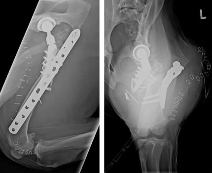
Faux profile and craniocadual projection of the left femur.
- There is a complete implant failure with displacement of the bony fragments and shortening
- Moderate accompanying soft tissue swelling
- The dog underwent a second revision
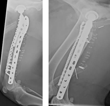
Faux profile of the left femur and lateral projection of the pelvis.
2 days later another radiographic study was performed.
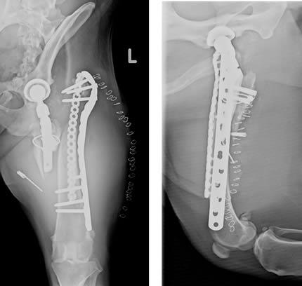
Craniocaudal view and faux profile of the left femur.
There was a new revision of the fracture with fixation as shown and removal of the implant on the proximal femur.

Craniocaudal view and faux profile of the left femur.
Another 2 days post OP radiographic examination was performed with similar results as before.
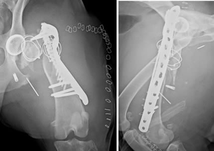
Craniocaudal view of the left femur and lateral view of the pelvis.
Followed by another revision 2 weeks later with graft and bone transplantation from a rib.

Craniocaudal view of the left femur and lateral view of the pelvis.
Comments
- During the second operation histological and bacteriological sample were taken
- The histological sample consisted with reactive scar tissue
- Contamination with a resistant strain of Staphylococcus pseuodointermedius was diagnosed


