Dog
Labrador Retriever
Female neutered
8 years of age
Suspicious of hit by car some hours before, not able to stand.
Lateral view of the abdomen and of the thorax were taken.
Radiographic examination

Left lateral projection of the abdomen.
Radiographic findings
- There is severe distraction with widening of the L5-6 intervertebral space, wedge shaped (arrow).
- Ventral subluxation of L6 with widening of the synovial joint space (arrow heads).
- Luxation of the left femoral head (empty arrow).
- Suspicious of reduced detail in the sublumbar area.
Radiographic examination

Close up of the left lateral view of the abdomen.
Radiographic diagnosis
- Fracture/Luxation L5-6 with suspected instability.
- Accompanying soft tissue swelling, bleeding.
- Luxation of the left femoral head.
- To further assess the lesion, a CT study was performed.
Radiographic findings

Close up of the CT study, bone window, multiplanar reconstruction (transverse plane on the left and dorsal plane on the right image) at the level of the L5-6 vertebra.
Radiographic diagnosis
- On CT the complete assessment of the dislocation was possible (not only ventral luxation but also lateral and rotational).
- There is mild incongruency of the right synovial joint (arrow).
- Additionally there are pin point mineral densities close to it as small bony fragments (arrow head).
- There was no fracture across the joints.
- The luxation was fixated as shown.
Comments

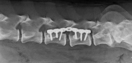
Ventrodorsal view (on the left ) and left lateral projection (on the right) of the lumbar spine, post surgery.
The femoral luxation was reduced in a second surgery.


